Premature Ovarian Failure (POF), also known as Hypergonadotropic Hypogonadism, refers to early depletion of ovarian follicles prior to age 40. Some definitions of POF are inclusive of patients with ovarian failure and primary amenorrhea, thus including patients with gonadal dysgenesis, while other definitions are limited to patients with secondary amenorrhea.
Approximately 1% of women will develop POF, however the incidence of POF is 10% to 28% in cases of primary amenorrhea. The diagnosis is based on amenorrhea with elevated serum FSH greater than 30 mIU/mL on at least two determinations.
Several etiologies of POF exist including: genetic, immune, enzymatic, ovarian trauma, and idiopathic. For most patients no specific cause can be identified, and their condition is classified as idiopathic. Recognizing that the normal physiology of the ovarian life Cycle involves gradual atresia, or loss of oocytes, in POF, the process is believed to result from an accelerated rate of atresia of the Oocyte pool.
Two intact X chromosomes are necessary for maintenance of oocytes during embryogenesis, and loss of or a structural abnormality in an X chromosome leads to accelerated oocyte loss. Defects in the X chromosome may result in various types of gonadal dysgenesis with variable times of onset of ovarian failure. Thus, even in Turner’s Syndrome (45,XO), or classic gonadal dysgenesis, some patients have been known to undergo spontaneous puberty and even onset of early menses prior to eventual secondary amenorrhea. These cases are a result of genetic mosaicism. However, even small defects in the X chromosome may be sufficient to cause POF.
A number of cases of ovarian failure have been reported with autoimmune disorders. While assays for detection of ovarian antibodies are becoming available, they are not yet widely used and/or standardized and so are of little clinical value at present.
From a practical standpoint, screening for the common autoimmune disorders is appropriate in women diagnosed with POF. Hypothyroidism is the autoimmune disorder most commonly seen in POF, and adrenal insufficiency due to adrenal antibodies is less common.
Under the category of enzymatic defects, galactosemia is a major cause of POF that is related to the toxic buildup of galactose in women who are unable to metabolize the sugar. Even in women with fairly well controlled galactose-free diets, POF tends to occur.
Another rare enzymatic defect linked to POF is 17 α-hydroxylase deficiency. Under the category of ovarian trauma, POF may be induced by ionizing radiation, chemotherapy, or overly aggressive ovarian surgery. Although not well documented, viral infections, particularly mumps, have been suggested to play a role in POF.
The management of premature ovarian failure will depend upon the age at which the syndrome is diagnosed. Evaluation of women under 30 who have POF should include a karyotype and screening for autoimmune disorders. A reasonable approach is to perform a few selected blood tests that should be obtained periodically as part of the long-term surveillance against associated autoimmune disorders.
These tests may include: TSH, Free T4, and subsequent thyroid antibodies if thyroid function is abnormal. Additionally, indications of adrenal disorders may be indicated by abnormal serum calcium, phosphorus, and fasting glucose. Aside from biochemical analysis, transvaginal ultrasound may be useful for assessing the size of the ovaries and the degree of follicular development, if any. In women diagnosed with onset of POF after age 30, the general consensus is that karyotype is not necessary.
Treatment usually consists of estrogen replacement. Because of their hypoestrogenic state, they are at increased risk for osteoporosis and cardiovascular disease and need appropriate surveillance and Hormone replacement. In young patients, this usually is given in the form of oral contraceptive pills that most often reinitiate normal menstrual cycling. Other variations of cyclic estrogen and progestin replacement are also feasible.
A patient who has been in ovarian failure for a number of years without treatment, regardless of age of onset, should have a baseline bone density determination and appropriate counseling regarding options for hormone replacement or other therapeutic interventions such as bisphosphonates.
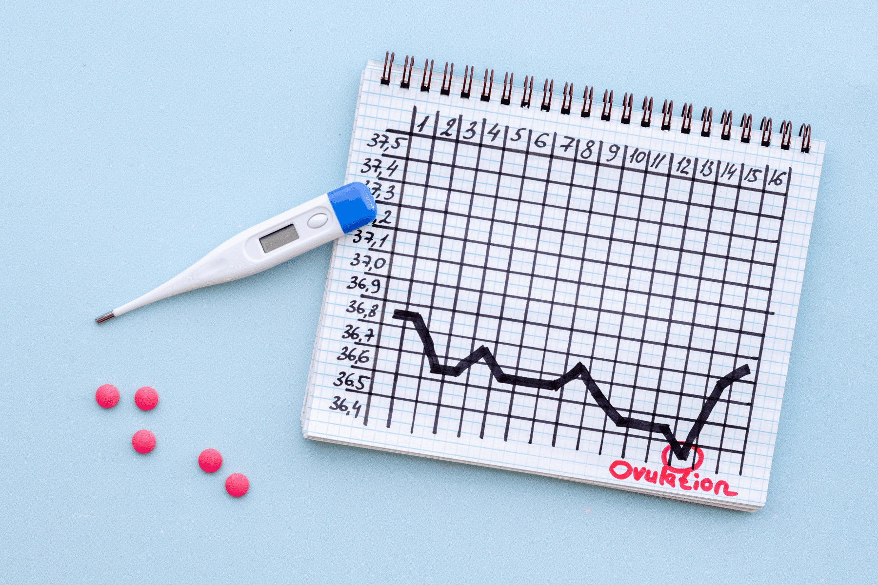
The best prospect for pregnancy is with egg donation, although pregnancy rates are reduced when using a sibling’s oocytes. Otherwise, pregnancy success with egg donation is approximately 60% per cycle if her partner has evidence of normal sperm and her uterine cavity is also normal. Sporadic and unpredictable Ovulation occurs in up to 25% of patients with POF who are continually monitored. However, the lifetime spontaneous pregnancy rate following this diagnosis is only 6-8%.
In the specific situation of Turner’s Syndrome, screening cardiac assessment is indicated prior to attempting pregnancy with egg donation. This is due to the fact that a recent literature review identified four case reports of Turner’s patients who died during pregnancy.
The maternal risk of death from rupture or dissection of the aorta in pregnancy may be 2% or higher. The recommendation prior to egg donation in Turner’s patients is echocardiography given the potential cardiac risk and its exacerbation from increased cardiac demand during pregnancy.
Medical contribution by Jane Nani, M.D.
Dr. Jane Nani is board certified in Obstetrics and Gynecology and in Reproductive Endocrinology and Infertility (REI), and has been practicing medicine since 1996.


